| BioMed: ★ An abdominal mass refers to an abnormal, space-occupying growth, the nature of which requires thorough evaluation to determine its
origin, composition, and clinical significance. These masses can arise from a wide range of causes, including congenital malformations, infections, benign or malignant neoplasms, hematologic disorders, vascular abnormalities such as aneurysms, and trauma. Systemic conditions like tuberculosis, lymphoma, or other inflammatory diseases may also present as abdominal masses. Such growths can displace, compress, infiltrate, or impair the function of nearby organs and structures, leading to symptoms such as pain, distention, intestinal obstruction, urinary retention, or weight loss, depending on their size and location.
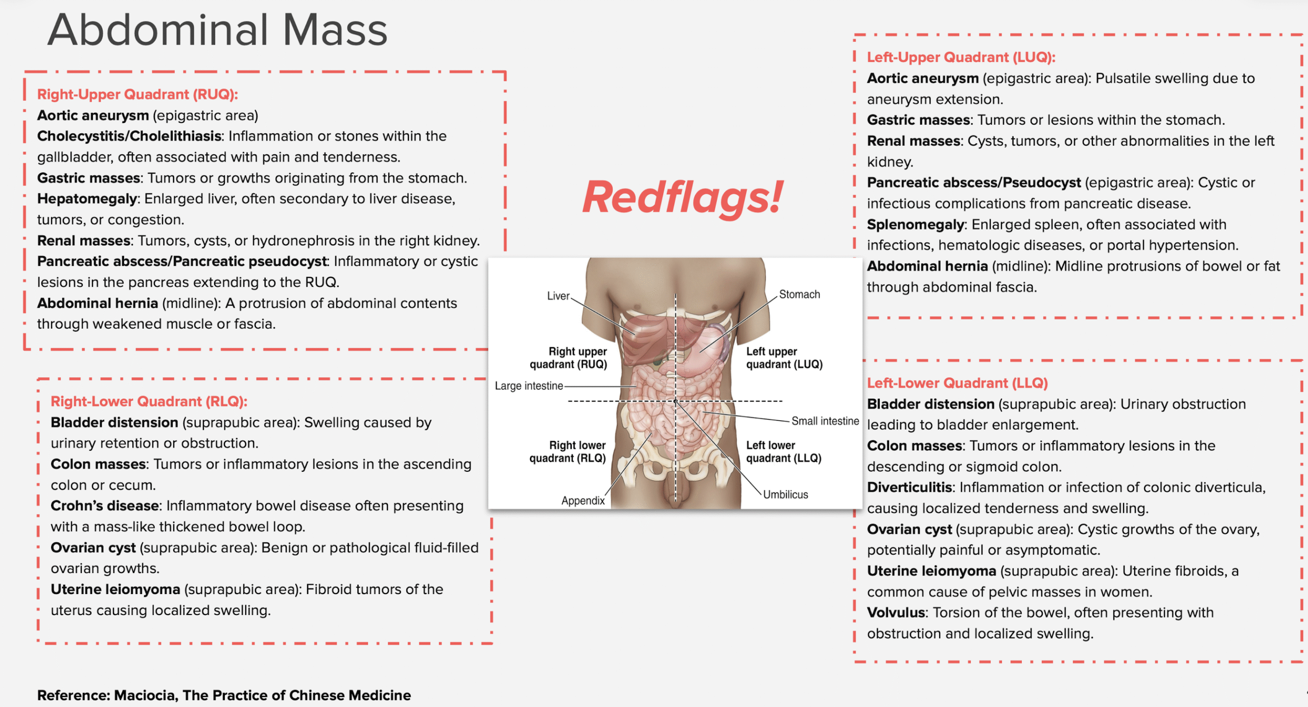
★ Abdominal masses can be categorized based on their anatomical location into three primary types:
1. Intraperitoneal – arising from structures within the peritoneal cavity such as the liver, spleen, gastrointestinal tract, or reproductive
organs.
2. Extraperitoneal – occurring outside the peritoneal cavity, either retroperitoneal (e.g., kidneys, adrenal glands, pancreas, lymph
nodes) or pelvic (e.g., reproductive organs, bladder, rectum).
3. Abdominal wall – originating from the musculature, fascia, or subcutaneous tissues of the abdominal wall.
★ Further diagnostic evaluation relies heavily on advanced imaging techniques, which play a pivotal role in determining the mass’s location,
size, composition, and relationship to adjacent structures. These techniques include:
1. Ultrasound (US): Often the initial imaging modality due to its non-invasiveness and ability to differentiate cystic from solid lesions.
2. US-guided core needle biopsy: Essential for obtaining tissue samples to confirm malignancy or infection.
3. CT scans with contrast: Provide detailed cross-sectional images, helping to identify vascular involvement, organ displacement, or
metastasis.
4. MRI: Particularly useful for soft tissue characterization, assessing pelvic masses, or distinguishing benign from malignant lesions. ○ PET scans: Used for evaluating metabolic activity, staging cancers, and detecting distant metastasis.
★ In some cases, additional laboratory tests, such as tumor markers (e.g., CA-125, AFP, CEA) or blood cultures, may be warranted to aid
diagnosis, especially when cancer, infection, or systemic conditions are suspected.
[34]
|
| Etiology: In the ancient text, abdominal masses are called jī jù (积聚). Ji indicates actual abdominal masses that are fixed and immovable; Ju indicates abdominal masses that come and go, do not have a fixed location and are movable. Another name for abdominal masses was zhēng jiǎ (症瘕), Zheng being equivalent to Ji (i.e. actual, fixed masses) and Jia being equivalent to Ju (i.e. non-substantial masses from stagnation of Qi). The term Zheng Jia normally referred to abdominal masses occurring only in women, but, although these masses are more frequent in women, they do occur in men as well. ★ Etiology: 1. Emotional strain: Prolonged emotional stress can disrupt the flow of Qi, leading to stagnation. 2. Irregular diet: Poor dietary habits can impair digestion, weaken the Spleen, and contribute to the formation of Phlegm or stagnation of Qi and Blood. 3. External pathogenic factors: External Cold can invade the lower abdomen, impairing the circulation of Blood and eventually
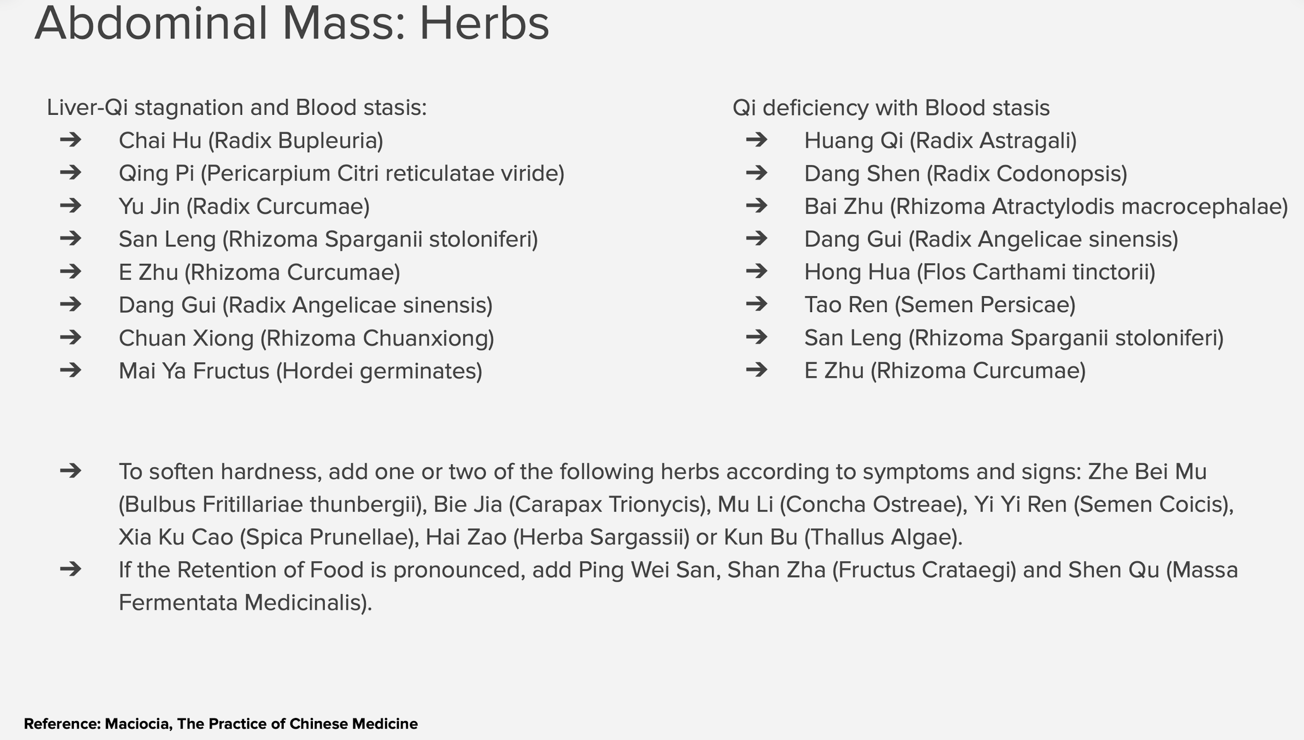
leading to Blood stasis. ★ Pathology: Abdominal masses are primarily characterized by Qi stagnation or Blood stasis. In some cases, the accumulation of Phlegm may also play a role. Importantly, there is always an underlying deficiency of Qi in patients with abdominal masses, which compromises the body’s ability to resolve the condition. ★ Masses from Qi stagnation: These masses are movable upon palpation, come and go, and may shift location. Qi stagnation leads to a temporary and less defined mass due to the disrupted but dynamic flow of Qi. ★ Masses from Blood stasis:These masses are fixed in location, hard to the touch, and non-movable on palpation.Blood stasis causes a dense and stagnant mass that remains unyielding and firmly rooted.
★ Masses from Phlegm: These masses are soft to the touch, have a fixed location, and are usually painless. The accumulation of Phlegm results in a softer texture, as Phlegm is a fluid and viscous pathogenic factor.
|